Applications of nanodiamonds in medical and sensor fields
-
摘要: 纳米金刚石不仅具有传统块状金刚石优异的物理和化学性质,还具有良好的生物相容性、光学特性和电学特性等。这些特性使得纳米金刚石在医疗和传感器领域的应用受到了科研人员的广泛关注。系统综述纳米金刚石的制备方法及各种方法的优缺点,指出其在表面改性、生物、光和电等方面的特殊性能,重点总结纳米金刚石在医疗和传感器领域近年来的研究,最后展望纳米金刚石未来的发展和应用前景,并指出其中的不足之处。Abstract:
Significance This study systematically reviews the applications of nanodiamonds in the medical and sensor industries, along with their fabrication techniques and inherent properties. Nanodiamonds, characterized by their unique physicochemical attributes, such as high hardness, favorable biocompatibility, optical characteristics, and electrical properties, are posited to hold substantial promise for diverse applications in these domains. Progress The article delineates a variety of synthesis methods for nanodiamonds, encompassing detonation synthesis, chemical vapor deposition, ball milling, high-temperature high-pressure synthesis, and laser ablation. It also presents an analytical review of the advantages and disadvantages inherent to each technique. Furthermore, the study addresses advancements in nanodiamond surface modification, biocompatibility, and electrical and optical properties. It concludes with a comprehensive summary of nanodiamond applications in the medical and sensor fields, highlighting their utilization in biological labeling and imaging, anti-infective therapy, tissue engineering and repair, cancer treatment, biosensors, electrochemical and gas sensors, and pressure sensors, among others. Conclusions and Prospects The article acknowledges the broad application prospects of nanodiamonds in the medical and sensor sectors, while also highlighting existing research gaps, such as the need for improved purity, yield, and size uniformity during synthesis, challenges in achieving efficient and controllable fluorescence for imaging, and an incomplete understanding of nanodiamonds' metabolic pathways and biological impact within living organisms. The article also speculates on potential future directions for nanodiamond research, including enhancing synthesis quality, achieving precise control over fluorescence properties, elucidating their metabolic pathways and biological effects, and developing more efficient and sensitive biosensors. This review article offers a comprehensive research perspective on the applications of nanodiamonds in the medical and sensor fields, and presents constructive suggestions for future research directions. -
Key words:
- nanodiamonds /
- performance /
- medical treatment /
- sensor
-
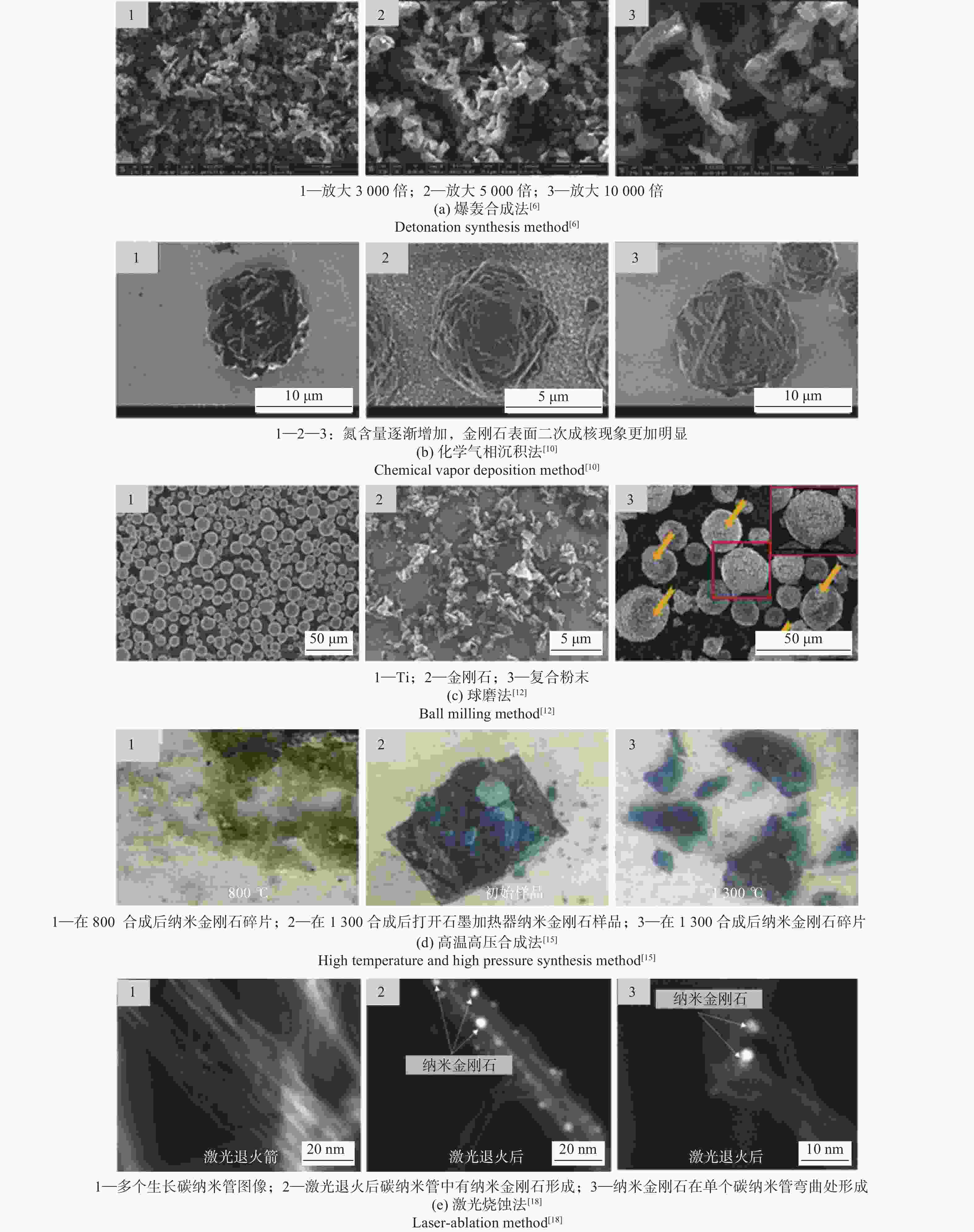
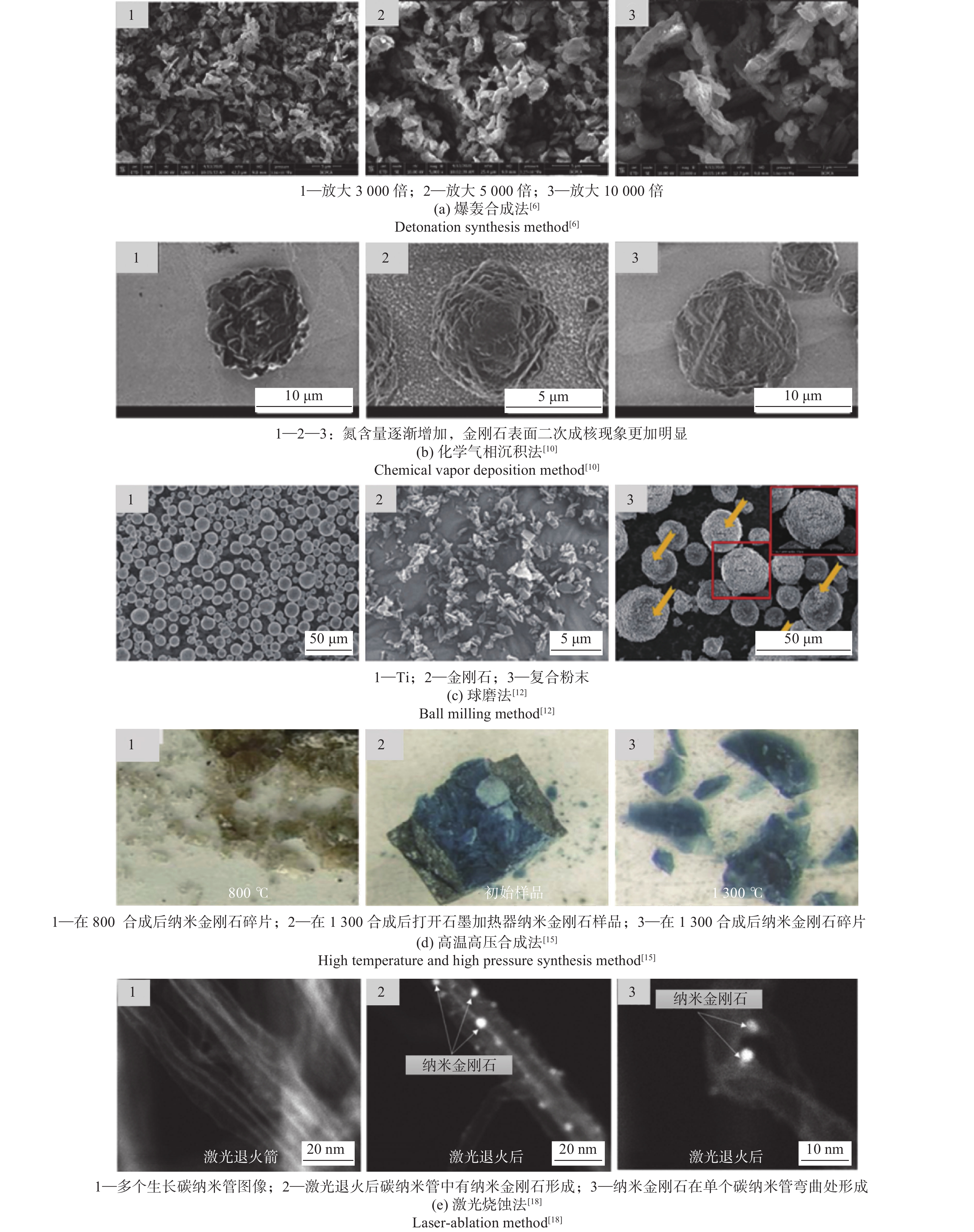
图 1 不同方法制备得到的纳米金刚石实物图
Figure 1. Physical images of nanodiamonds prepared by different methods
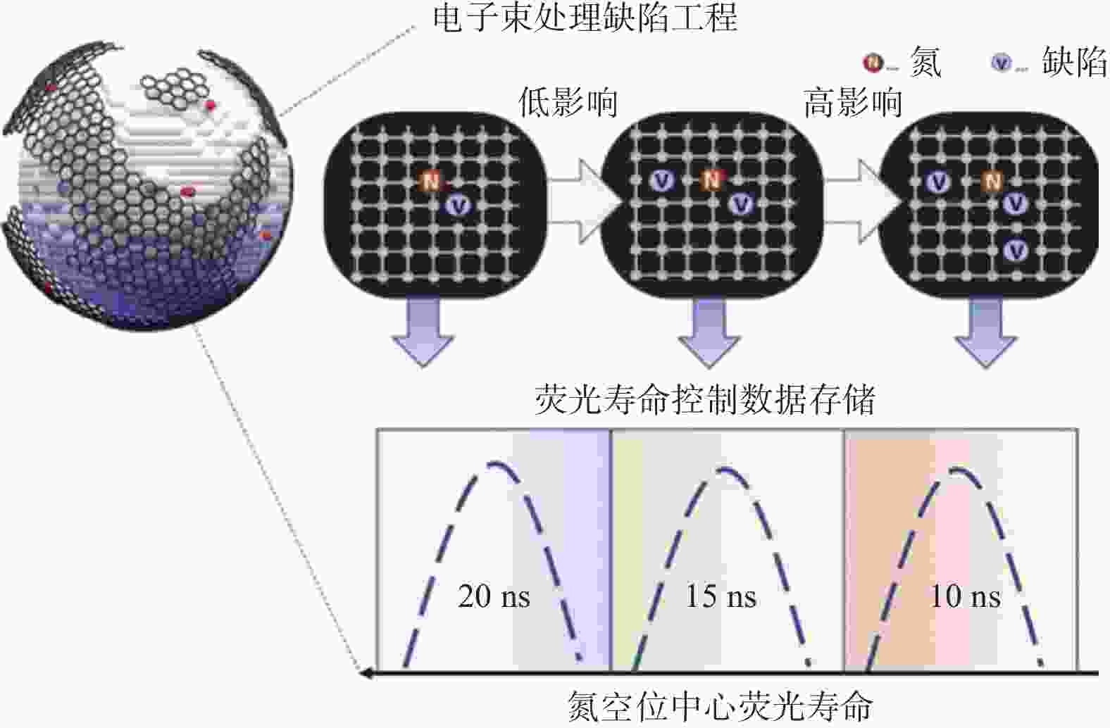

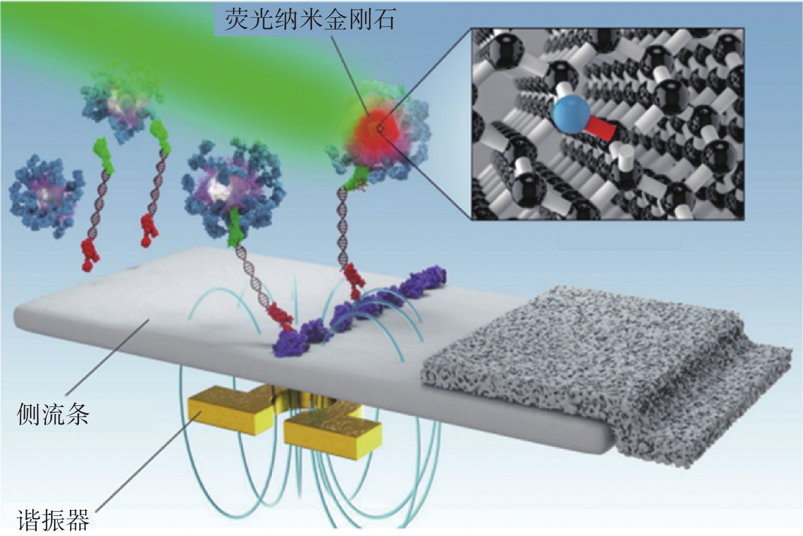
图 6 纳米金刚石在荧光成像领域的应用
Figure 6. Application of nanodiamonds in fluorescence imaging field
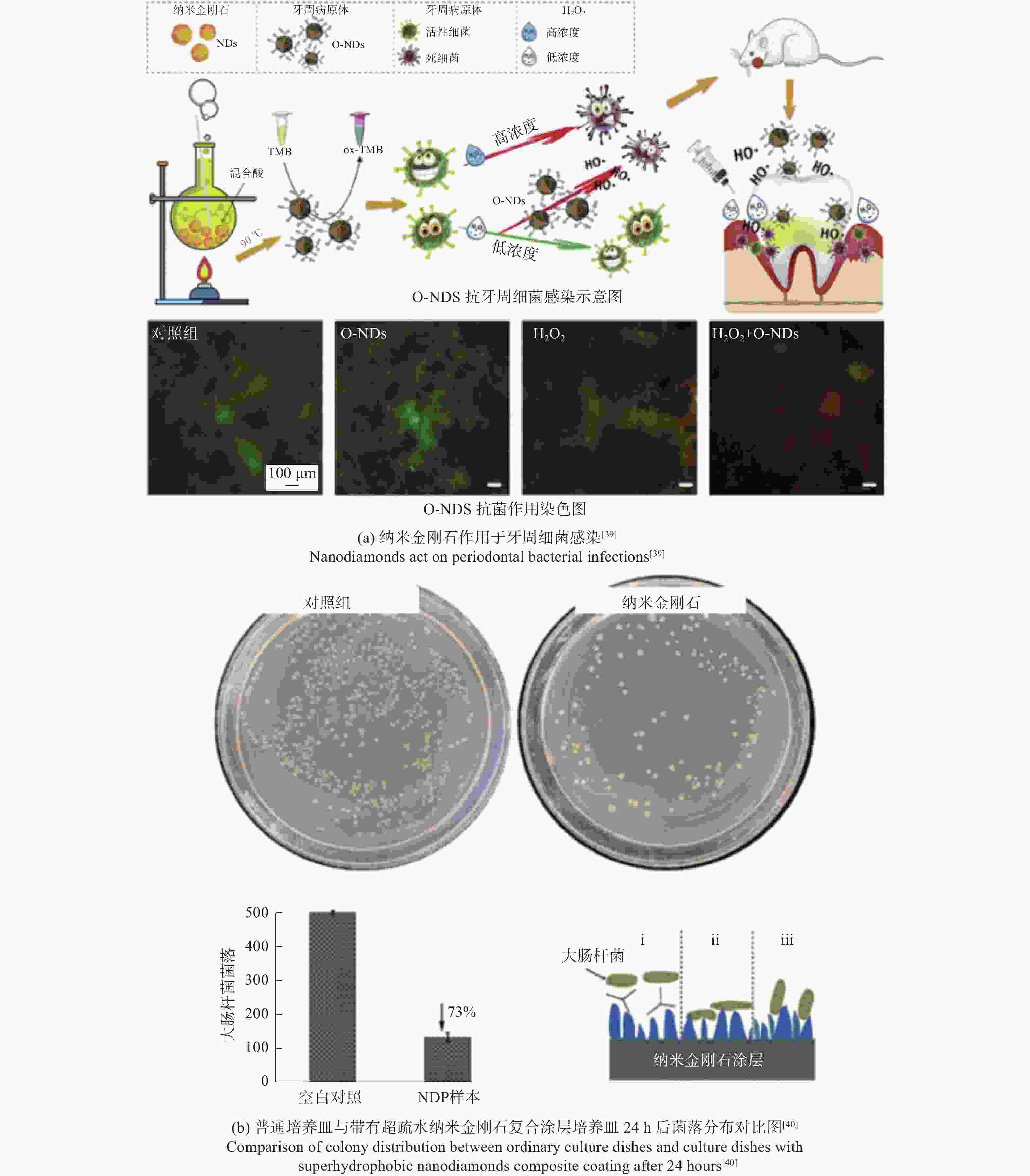
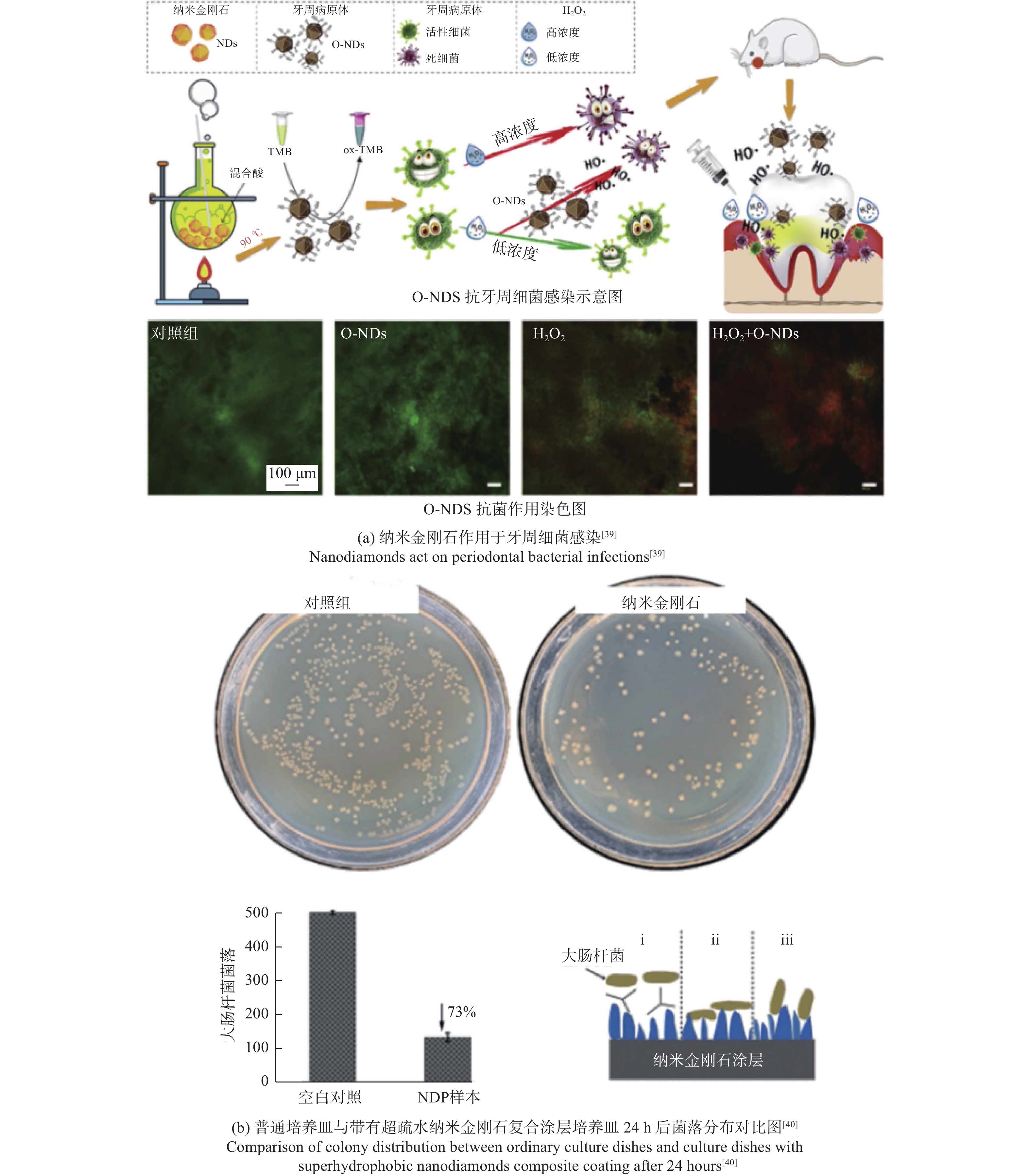
图 7 纳米金刚石在抗菌和抗感染领域的应用
Figure 7. Application of nanodiamonds in antibacterial and anti infective fields
表 1 纳米金刚石的制备方法、优缺点和未来研究重点
Table 1. Preparation methods, advantages and disadvantages, and future research focus of nanodiamonds
制备方法 粒径 d / nm 优缺点 未来研究重点 爆轰合成法[5-8] 2~200 优点:高效快速、工艺简单、尺寸范围广;
缺点:团聚现象严重、能耗较高、杂质较多更精确地控制爆轰合成的条件,包括爆炸能量、温度、压力和反应时间等;探索团聚现象的解决方案 化学气相沉积法[9-10] 10~100 优点:尺寸均匀性好、制备效率高、制备形态丰富
(颗粒、薄膜等);
缺点:能耗高、催化剂成本昂贵、废气处理烦琐更精确地控制化学气相沉积法中的生长条件,包括气相组成、温度、压力和沉积时间等;探索新型前驱体 球磨法[11-13] 5~500 优点:尺寸范围广、设备简单、适用于多种碳源;
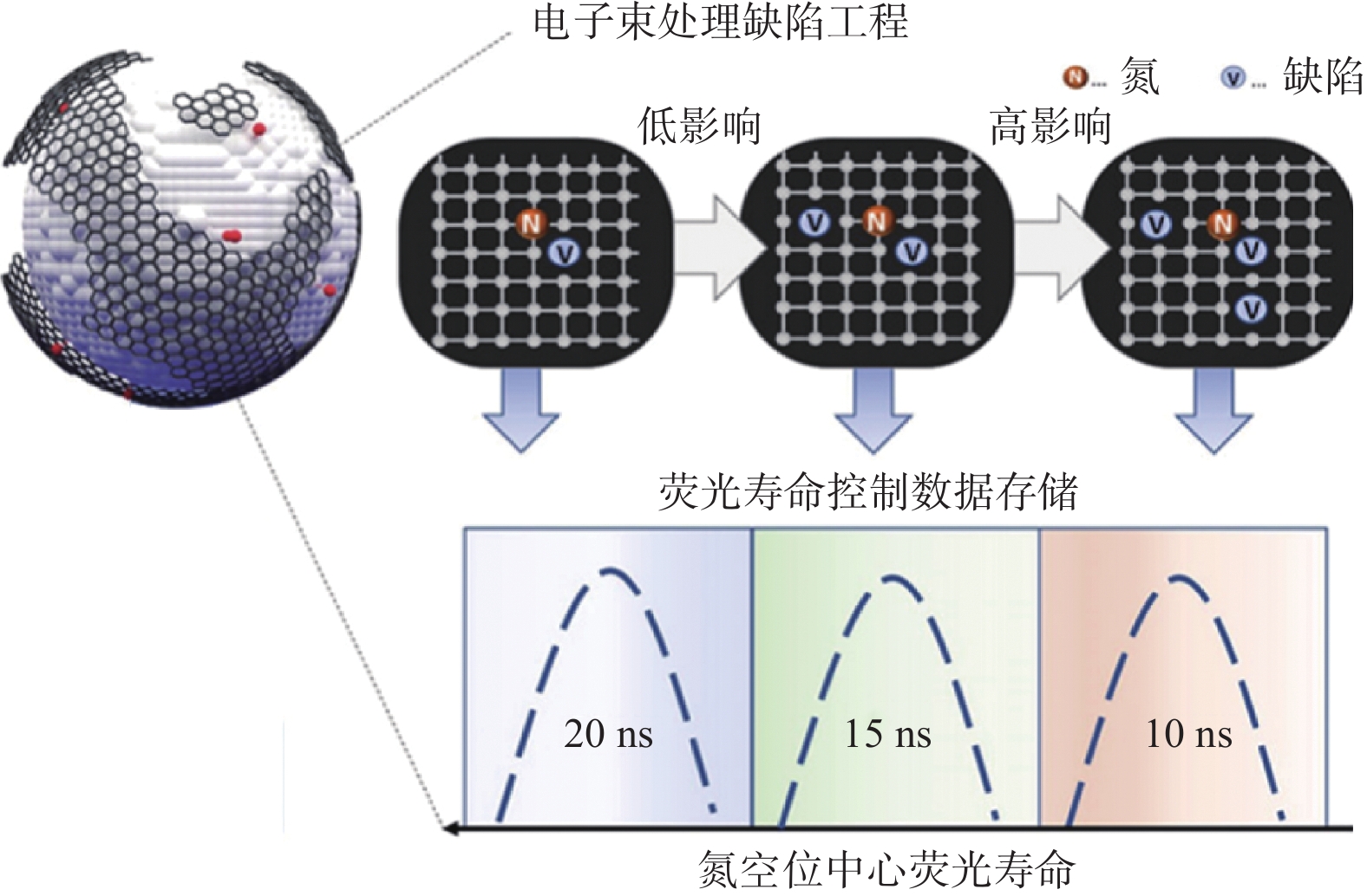
缺点:表面损伤、形貌不可控、杂质较多进一步优化球磨的条件,包括球磨时间、碾磨介质、碾磨速度等;探索新型碳源 高温高压合成法[14-16] 20~60 优点:尺寸可控、纯度高、适用于大规模生产;
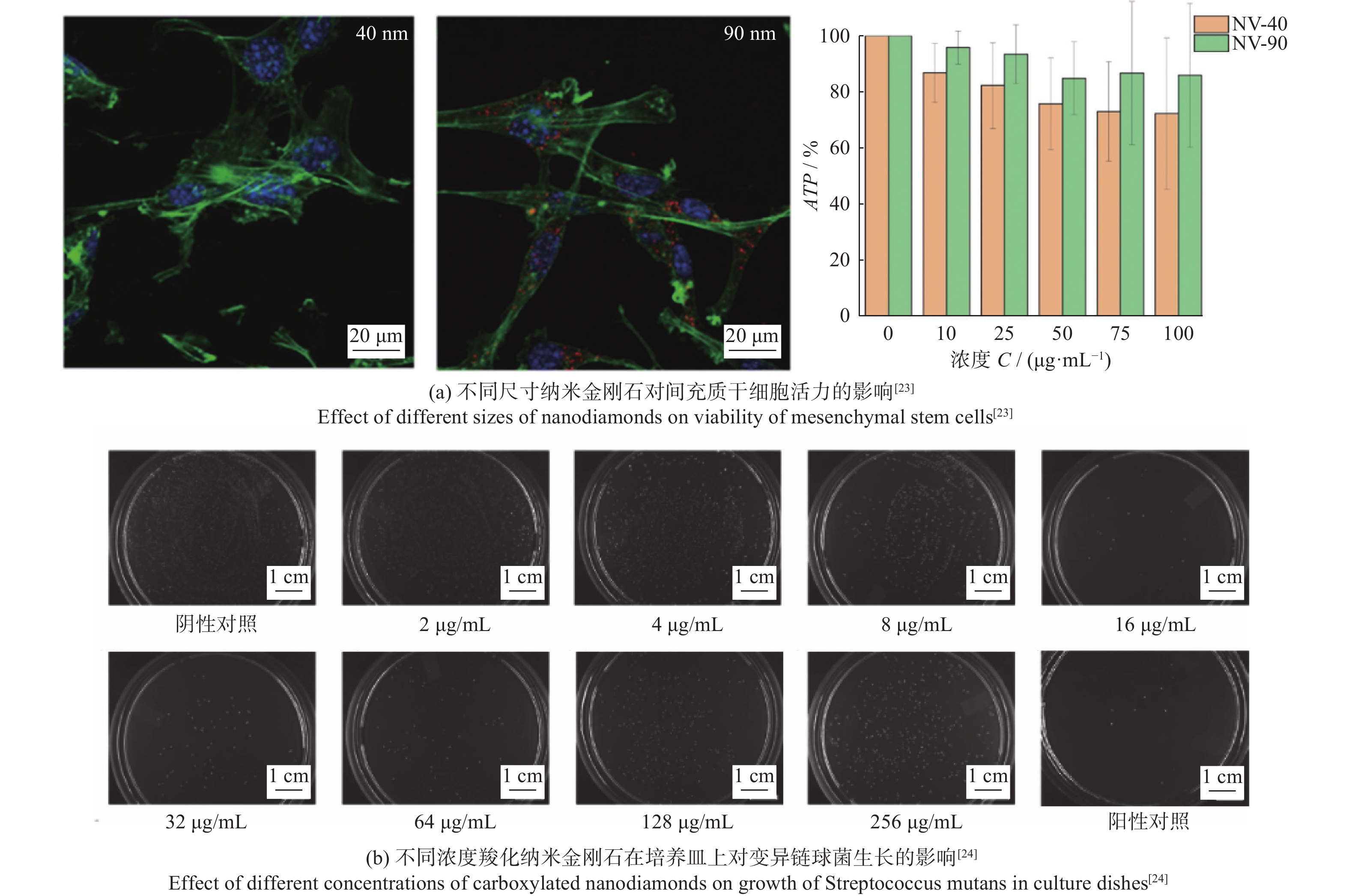
缺点:能耗高、设备昂贵、制备时间长更精确地控制合成条件;探索新型催化剂、碳源 激光烧蚀法[17-18] 2~10 优点:效率高、安全性高、可控性高、无须催化剂;
缺点:表面损伤、光源要求高、制备环境要求严苛进一步优化激光参数,包括激光功率、脉冲宽度、频率等;探索和应用新型激光源  下载: 导出CSV
表 2 纳米金刚石在医疗和传感器领域的应用与优势
Table 2. Application and advantages of nanodiamonds in medical and sensor fields
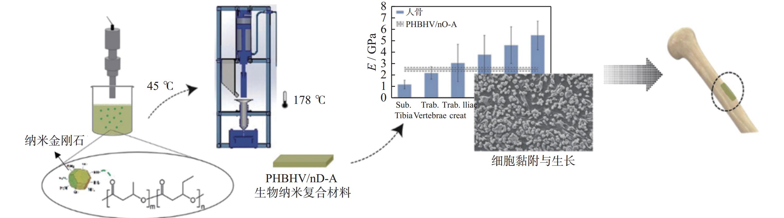
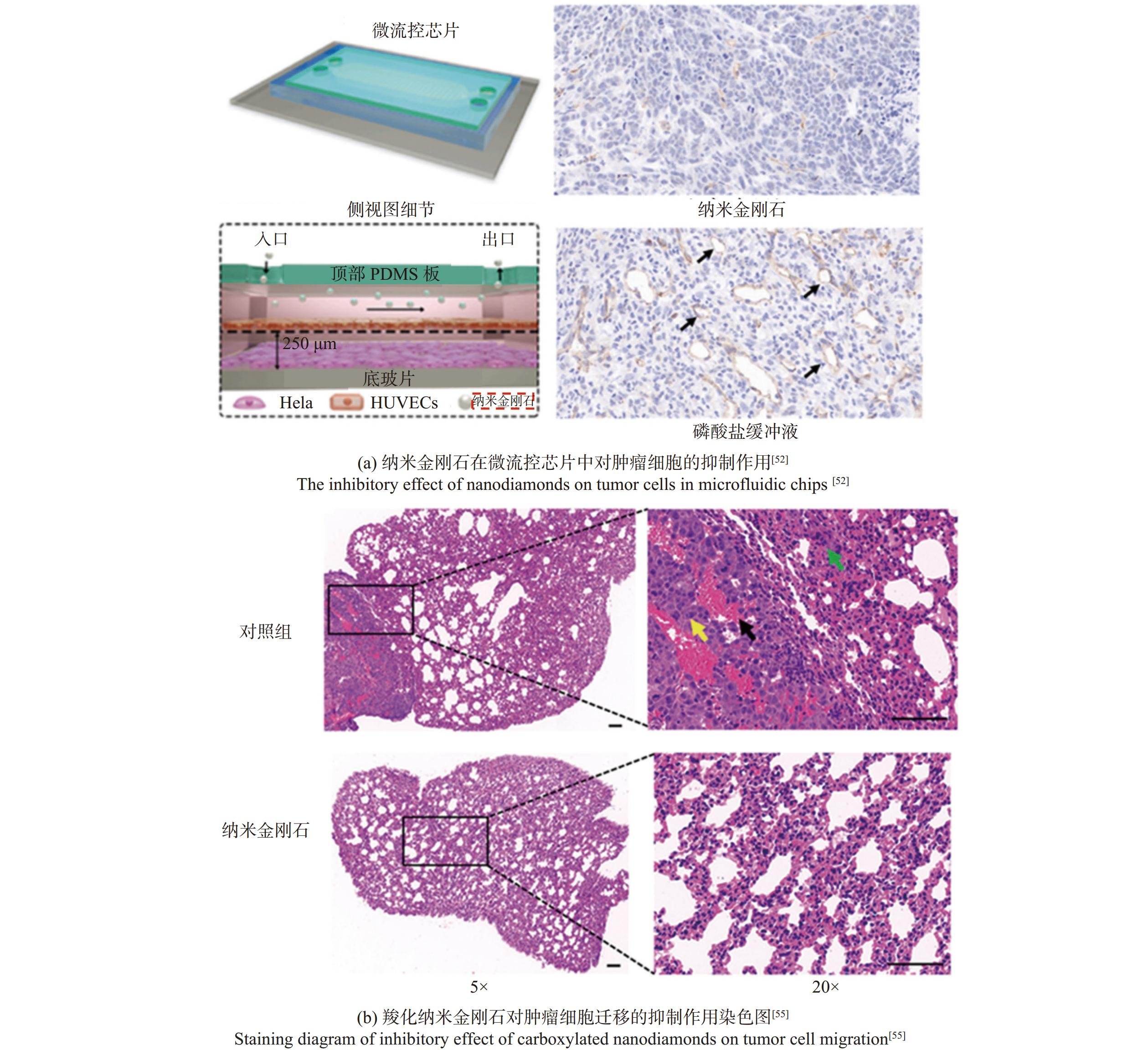
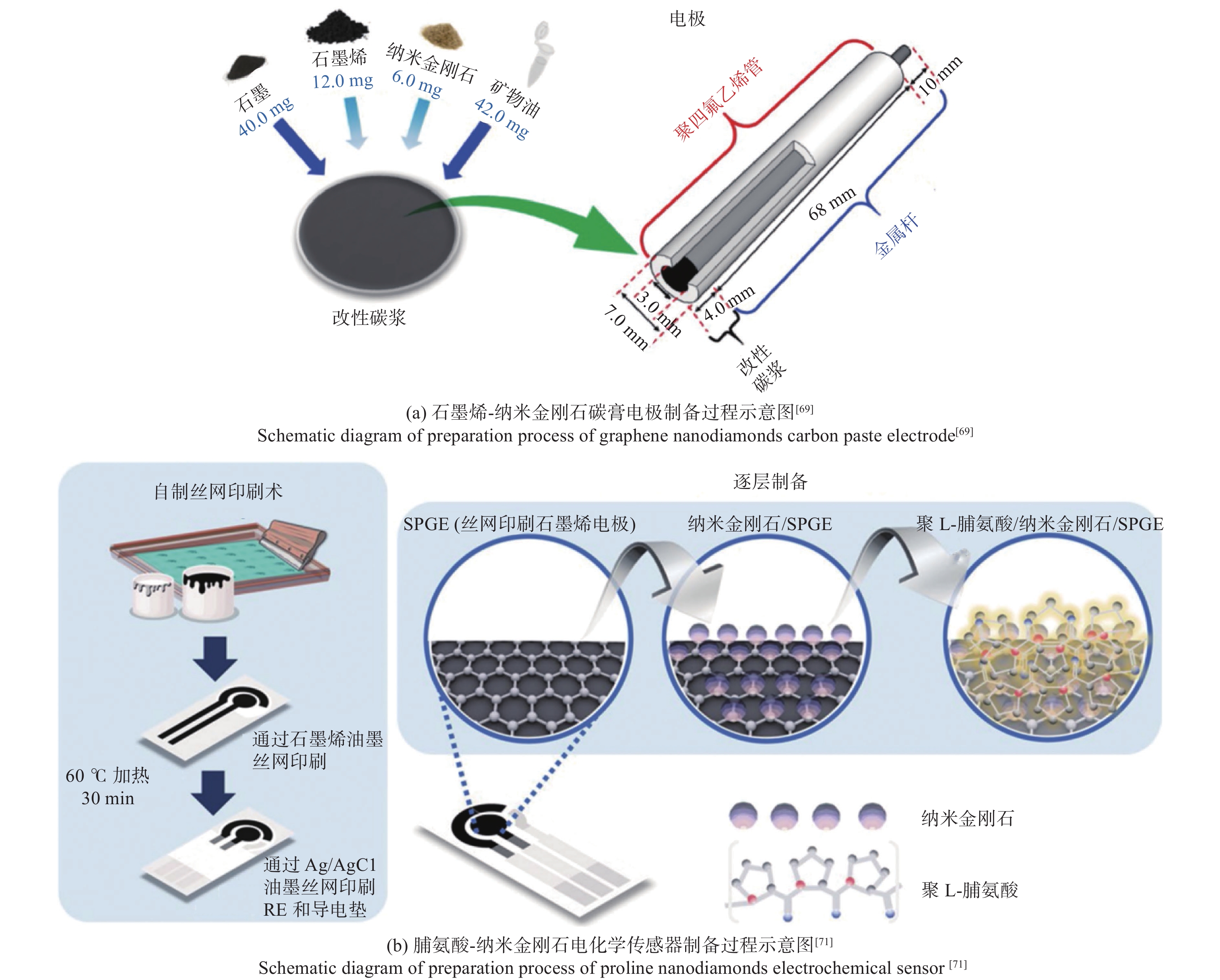
应用领域 特性 优势 生物标记与荧光成像 生物相容性、化学稳定性、导电性、荧光特性 精准捕捉、高敏感性、高分辨率 抗菌与抗感染治疗 生物相容性、高比表面积、稳定的晶格结构 抗菌、抗感染、物理破坏、氧化性 组织工程与修复 优异的机械性能、化学稳定性、生物相容性 精确控制和刺激、增强力学性能、结构复杂多样 癌症治疗 表面改性、黏附性、抑制癌细胞扩散游离特性 精准靶向治疗、穿透性强、传递效果好 生物传感器 生物相容性、生物惰性、化学稳定性、荧光特性 高灵敏性、高精度、高效选择性 电化学和气体传感器 大比表面积、导电性、优异的机械性能 强捕捉能力、快速检测能力、高灵敏性、高效选择性 压力传感器 高硬度、结构稳定性、优异的机械性能 高耐用性和耐磨性、高精准性、优异导热性
下载: 导出CSV
-
[1] AUCIELLO O, ASLAM D M. Review on advances in microcrystalline, nanocrystalline and ultrananocrystalline diamond films-based micro/nano-electromechanical systems technologies [J]. Journal of Materials Science,2021,56(12):7171-7230. doi: 10.1007/s10853-020-05699-9 [2] SKOOG S A, KUMAR G, ZHENG J W, et al. Biological evaluation of ultrananocrystalline and nanocrystalline diamond coatings [J]. Journal of Materials Science Materials in Medicine,2016,27(12):187. doi: 10.1007/s10856-016-5798-y [3] QIN J X, YANG X G, LV C F, et al. Nanodiamonds: Synthesis, properties, and applications in nanomedicine [J]. Materials & Design,2021,210:110091. doi: 10.1016/j.matdes.2021.110091 [4] 罗珊, 胡小月, 王成勇, 等. 金刚石在医疗领域的应用 [J]. 金刚石与磨料磨具工程,2018,38(2):1-7. doi: 10.13394/j.cnki.jgszz.2018.2.0001LUO Shan, HU Xiaoyue, WANG Chengyong, et al. Diamond application in medical field [J]. Diamond & Abrasives Engineering,2018,38(2):1-7. doi: 10.13394/j.cnki.jgszz.2018.2.0001 [5] DECARLI P S, JAMIESON J C. Formation of diamond by explosive shock [J]. Science,1961,133(3467):1821-1822. doi: 10.1126/science.133.3467.1821 [6] SHANG S Y, TONG Y, WANG Z C, et al. Study on the polycrystalline mechanism of polycrystalline diamond synthesized from graphite by direct detonation method [J]. Materials,2022,15(12):4154. doi: 10.3390/ma15124154 [7] YAN X R, LI X J, WANG X H, et al. Synthesis of nano-diamond/alumina composite by detonation method [J]. Diamond and Related Materials,2017,77:79-83. doi: 10.1016/j.diamond.2017.05.015 [8] 姚凯丽, 代兵, 乔鹏飞, 等. 纳米金刚石材料的研究进展 [J]. 人工晶体学报,2019,48(11):1977-1989. doi: 10.3969/j.issn.1000-985X.2019.11.001YAO Kaili, DAI Bing, QIAO Pengfei, et al. Research progress of nano-diamond materials [J]. Journal of Synthetic Crystals,2019,48(11):1977-1989. doi: 10.3969/j.issn.1000-985X.2019.11.001 [9] 徐帅, 李晓普, 丁玉龙, 等. 化学气相沉积金刚石微球的生长机制研究 [J]. 金刚石与磨料磨具工程,2018,38(5):1-5. doi: 10.13394/j.cnki.jgszz.2018.5.0001XU Shuai, LI Xiaopu, DING Yulong, et al. Chemical vapor deposition of diamond microsphere: Investigation of growth dynamics [J]. Diamond & Abrasives Engineering,2018,38(5):1-5. doi: 10.13394/j.cnki.jgszz.2018.5.0001 [10] ZHENG S J, WANG Q, GUO K S, et al. Effect of plasma modulation on the nucleation and crystal evolution of nanodiamond seeds during CVD diamond growth [J]. Journal of Crystal Growth,2024,627:127538. doi: 10.1016/j.jcrysgro.2023.127538 [11] EL-ESKANDARANY M S. Mechanically induced graphite-nanodiamonds-phase transformations during high-energy ball milling [J]. Journal of Materials Engineering and Performance,2017,26(6):2974-2982. doi: 10.1007/s11665-017-2711-x [12] LI W Q, ZHOU H X, LI X T, et al. Preparation and tribological properties of novel cold–sprayed Ti–diamond composite coating [J]. Surface and Coatings Technology,2024,477:130386. doi: 10.1016/j.surfcoat.2024.130386 [13] KATZENSTEINER A, ROSALIE J M, PIPPAN R, et al. Synthesis of nanodiamond reinforced silver matrix nanocomposites: Microstructure and mechanical properties [J]. Materials Science and Engineering: A,2020,782:139254. doi: 10.1016/j.msea.2020.139254 [14] HALL H T. The synthesis of diamond [J]. Journal of Chemical Education,1961,38(10):484. doi: 10.1021/ed038p484 [15] EKIMOV E, SHIRYAEV A A, GRIGORIEV Y, et al. Size-dependent thermal stability and optical properties of ultra-small nanodiamonds synthesized under high pressure [J]. Nanomaterials,2022,12(3):351. doi: 10.3390/nano12030351 [16] EKIMOV E A, SHIRYAEV A A, SIDOROV V A, et al. Synthesis and properties of nanodiamonds produced by HPHT carbonization of 1-fluoroadamantane [J]. Diamond and Related Materials,2023,136:109907. doi: 10.1016/j.diamond.2023.109907 [17] YOGESH G K, SHUKLA S, SASTIKUMAR D, et al. Progress in pulsed laser ablation in liquid (PLAL) technique for the synthesis of carbon nanomaterials: A review [J]. Applied Physics A,2021,127(11):810. doi: 10.1007/s00339-021-04951-6 [18] HAQUE A, SACHAN R, NARAYAN J. Synthesis of diamond nanostructures from carbon nanotube and formation of diamond-CNT hybrid structures [J]. Carbon,2019,150:388-395. doi: 10.1016/j.carbon.2019.05.027 [19] TEGAFAW T, LIU S W, AHMAD M Y, et al. Production, surface modification, physicochemical properties, biocompatibility, and bioimaging applications of nanodiamonds [J]. RSC Advances,2023,13(46):32381-32397. doi: 10.1039/d3ra06837d [20] YANG N J, YU S Y, MACPHERSON J V, et al. Conductive diamond: Synthesis, properties, and electrochemical applications [J]. Chemical Society Reviews,2019,48(1):157-204. doi: 10.1039/C7CS00757D [21] PETRONE A, BECK R A, KASPER J M, et al. Electronic structures and spectroscopic signatures of silicon-vacancy containing nanodiamonds [J]. Physical Review B,2018,98(20):205405. doi: 10.1103/PhysRevB.98.205405 [22] LAUBE C, TEMME R, PRAGER A, et al. Fluorescence lifetime control of nitrogen vacancy centers in nanodiamonds for long-term information storage [J]. ACS Nano,2023,17(16):15401-15410. doi: 10.1021/acsnano.3c00328 [23] FRYER C, MURRAY P, ZHANG H F. Evaluation of cytotoxicity and bioimaging of nitrogen-vacancy nanodiamonds [J]. Nanomaterials,2022,12(23):4196. doi: 10.3390/nano12234196 [24] QUAN C T, LIN H Y, XIAO H, et al. Inhibitory effect of carboxylated nanodiamond on oral pathogenic bacteria streptococcus mutans [J]. Journal of Clinical Laboratory Analysis,2021,35(8):e23872. doi: 10.1002/jcla.23872 [25] LI D D, CHEN X, WANG H, et al. Cetuximab-conjugated nanodiamonds drug delivery system for enhanced targeting therapy and 3D Raman imaging [J]. Journal of Biophotonics,2017,10(12):1636-1646. doi: 10.1002/jbio.201700011 [26] LI D D, CHEN X, WANG H, et al. Nanodiamonds as Raman probes for specifically targeted bioimaging: Visualization and mechanism study of the biorecognition between nanodiamonds-EGF and EGFR [J]. RSC Advances,2017,7(21):12835-12841. doi: 10.1039/C6RA28139G [27] LAUBE C, RIYAD Y M, LOTNYK A, et al. Defined functionality and increased luminescence of nanodiamonds for sensing and diagnostic applications by targeted high temperature reactions and electron beam irradiation [J]. Materials Chemistry Frontiers,2017,1(12):2527-2540. doi: 10.1039/C7QM00241F [28] SIMPSON D A, MORRISROE E, MCCOEY J M, et al. Non-neurotoxic nanodiamond probes for intraneuronal temperature mapping [J]. ACS Nano,2017,11(12):12077-12086. doi: 10.1021/acsnano.7b04850 [29] IKLIPTIKAWATI D K, HAZAWA M, SO F T K, et al. Label-free tomographic imaging of nanodiamonds in living cells [J]. Diamond and Related Materials,2021,118:108517. doi: 10.1016/j.diamond.2021.108517 [30] SELVAM R, GANDHI A, HUNG S C, et al. Multifunctional ferromagnetic nanodiamond for dual-mode fluorescence imaging and magnetic drug targeting [J]. Diamond and Related Materials,2023,139:110398. doi: 10.1016/j.diamond.2023.110398 [31] FARD M G, KHABIR Z, REINECK P, et al. Targeting cell surface glycans with lectin-coated fluorescent nanodiamonds [J]. Nanoscale Advances,2022,4(6):1551-1564. doi: 10.1039/D2NA00036A [32] YANAGI T, KAMINAGA K, SUZUKI M, et al. All-optical wide-field selective imaging of fluorescent nanodiamonds in cells, in vivo and ex vivo [J]. ACS Nano,2021,15(8):12869-12879. doi: 10.1021/acsnano.0c07740 [33] LAPORTE G, PSALTIS D. STED imaging of green fluorescent nanodiamonds containing nitrogen-vacancy-nitrogen centers [J]. Biomedical Optics Express,2015,7(1):34-44. doi: 10.1364/BOE.7.000034 [34] TERADA D, SEGAWA T F, SHAMES A I, et al. Monodisperse five-nanometer-sized detonation nanodiamonds enriched in nitrogen-vacancy centers [J]. ACS Nano,2019,13(6):6461-6468. doi: 10.1021/acsnano.8b09383 [35] WEHLING J, DRINGEN R, ZARE R N, et al. Bactericidal activity of partially oxidized nanodiamonds [J]. ACS Nano,2014,8(6):6475-6483. doi: 10.1021/nn502230m [36] ONG S Y, HARMELEN R J J V, NOROUZI N, et al. Interaction of nanodiamonds with bacteria [J]. Nanoscale,2018,10(36):17117-17124. doi: 10.1039/C8NR05183F [37] CHEN M, LI Y, HOU W X, et al. The antibacterial effect, biocompatibility, and osteogenesis of vancomycin-nanodiamond composite scaffold for infected bone defects [J]. International Journal of Nanomedicine,2023,18:1365-1380. doi: 10.2147/IJN.S397316 [38] GUTIÉRREZ B J M, CONCEIÇÃO K, DE ANDRADE V M, et al. High antibacterial properties of DLC film doped with nanodiamond [J]. Surface and Coatings Technology,2019,375:395-401. doi: 10.1016/j.surfcoat.2019.07.029 [39] FANG J, WANG H, BAO X F, et al. Nanodiamond as efficient peroxidase mimic against periodontal bacterial infection [J]. Carbon,2020,169:370-381. doi: 10.1016/j.carbon.2020.07.055 [40] UZOMA P C, WANG Q M, ZHANG W Y, et al. Anti-bacterial, icephobic, and corrosion protection potentials of superhydrophobic nanodiamond composite coating [J]. Colloids and Surfaces A: Physicochemical and Engineering Aspects,2021,630:127532. doi: 10.1016/j.colsurfa.2021.127532 [41] CHANG B M, PAN L, LIN H H, et al. Nanodiamond-supported silver nanoparticles as potent and safe antibacterial agents [J]. Scientific Reports,2019,9(1):13164. doi: 10.1038/s41598-019-49675-z [42] RIBEIRO M E A, CHECCA HUAMAN N R, GOMEZ J G C, et al. Poly(3-hydroxybutyrate-co-3-hydroxyvalerate) and amino-functionalized nanodiamond bionanocomposites for bone tissue defect repair [J]. International Journal of Biological Macromolecules,2023,226:1041-1053. doi: 10.1016/j.ijbiomac.2022.11.221 [43] STIGLER R G, SCHIMKE M M, BIGUS S, et al. Pervasion of beta-tricalcium phosphate with nanodiamond particles yields efficient and safe bone replacement material amenable for biofunctionalization and application in large-size osseous defect healing [J]. Nanomedicine: Nanotechnology, Biology and Medicine,2019,16:250-257. doi: 10.1016/j.nano.2018.08.015 [44] CHOI S, NOH S H, LIM C O, et al. Icariin-functionalized nanodiamonds to enhance osteogenic capacity in vitro [J]. Nanomaterials,2020,10(10):2071. doi: 10.3390/nano10102071 [45] SHUAI C J, LI Y, WANG G Y, et al. Surface modification of nanodiamond: Toward the dispersion of reinforced phase in poly-L-lactic acid scaffolds [J]. International Journal of Biological Macromolecules,2019,126:1116-1124. doi: 10.1016/j.ijbiomac.2019.01.004 [46] WANG L N, CAO W W, WANG X, et al. Biodegradable silver-loaded polycation modified nanodiamonds /polyurethane scaffold with improved antibacterial and mechanical properties for cartilage tissue repairing [J]. Journal of Materials Science Materials in Medicine,2019,30(4):41. doi: 10.1007/s10856-019-6244-8 [47] SHUAI C J, HUANG W, FENG P, et al. Nanodiamond reinforced polyvinylidene fluoride/bioglass scaffolds for bone tissue engineering [J]. Journal of Porous Materials,2017,24(1):249-255. doi: 10.1007/s10934-016-0258-0 [48] GUPTA S K, TRIPATHY S P, BHARTI D, et al. One pot synthesis of phosphate glass with in situ formed nanodiamonds from adenosine triphosphate for bone repair [J]. Ceramics International,2023,49(13):22537-22546. doi: 10.1016/j.ceramint.2023.04.089 [49] IBRAHIM M, XUE Y, OSTERMANN M, et al. In vitro cytotoxicity assessment of nanodiamond particles and their osteogenic potential [J]. Journal of Biomedical Materials Research Part A,2018,106(6):1697-1707. doi: 10.1002/jbm.a.36369 [50] GU M J, WANG X, TOH T B, et al. Nanodiamond-based platform for intracellular-specific delivery of therapeutic peptides against hepatocellular carcinoma [J]. Advanced Therapeutics,2018,1(8):1800110. doi: 10.1002/adtp.201800110 [51] XU J R, GU M J, HOOI L, et al. Enhanced penetrative siRNA delivery by a nanodiamond drug delivery platform against hepatocellular carcinoma 3D models [J]. Nanoscale,2021,13(38):16131-16145. doi: 10.1039/d1nr03502a [52] GUO Q Y, LI L, GAO G Y, et al. Nanodiamonds inhibit the proliferation and migration of endothelial cells in a tumor/endothelial cells co-culture microfluidic system [J]. Carbon,2024,218:118671. doi: 10.1016/j.carbon.2023.118671 [53] NISHIKAWA M, KANG H G, ZOU Y J, et al. Conjugation of phenylboronic acid moiety through multistep organic transformations on nanodiamond surface for an anticancer nanodrug for boron neutron capture therapy [J]. Bulletin of the Chemical Society of Japan,2021,94(9):2302-2312. doi: 10.1246/bcsj.20210200 [54] YOSHINO F, AMANO T, ZOU Y J, et al. Preferential tumor accumulation of polyglycerol functionalized nanodiamond conjugated with cyanine dye leading to near-infrared fluorescence in vivo tumor imaging [J]. Small,2019,15(48):e1901930. doi: 10.1002/smll.201901930 [55] GUO Q Y, LI L, GAO G Y, et al. Nanodiamonds inhibit cancer cell migration by strengthening cell adhesion: Implications for cancer treatment [J]. ACS Applied Materials & Interfaces,2021,13(8):9620-9629. doi: 10.1039/D1NR03502A [56] CUI Z F, ZHANG Y, XIA K, et al. Nanodiamond autophagy inhibitor allosterically improves the arsenical-based therapy of solid tumors [J]. Nature Communications,2018,9(1):4347. doi: 10.1038/s41467-018-06749-2 [57] ACUÑA-AGUILAR L E, CONEJO-DÁVILA A S, MIKI-YOSHIDA M, et al. The improved cytotoxic capacity of functionalized nanodiamonds with metformin in breast and ovarian cancer cell lines [J]. Processes,2023,11(9):2616. doi: 10.3390/pr11092616 [58] NAZARKOVSKY M, DE MELLO H L, BISAGGIO R C, et al. Hybrid suspension of nanodiamonds-nanosilica/titania in cytotoxicity tests on cancer cell lines [J]. Inorganic Chemistry Communications,2020,111:107673. doi: 10.1016/j.inoche.2019.107673 [59] WEI W H W, FADHILAH G, LEE C C, et al. Nanomaterial-based biosensors for avian influenza virus: A new way forward [J]. Talanta,2023,265:124892. doi: 10.1016/j.talanta.2023.124892 [60] XU J R, ZHENG M J, THNG D K H, et al. NanoBeacon. AI: AI-enhanced nanodiamond biosensor for automated sensitivity prediction to oxidative phosphorylation inhibitors [J]. ACS Sensors,2023,8(5):1989-1999. doi: 10.1021/acssensors.3c00126 [61] MILLER B S, BEZINGE L, GLIDDON H D, et al. Spin-enhanced nanodiamond biosensing for ultrasensitive diagnostics [J]. Nature,2020,587(7835):588-593. doi: 10.1038/s41586-020-2917-1 [62] KAÇAR C, ERDEN P E. An amperometric biosensor based on poly(l-aspartic acid), nanodiamond particles, carbon nanofiber, and ascorbate oxidase–modified glassy carbon electrode for the determination of l-ascorbic acid [J]. Analytical and Bioanalytical Chemistry,2020,412(22):5315-5327. doi: 10.1007/s00216-020-02747-w [63] VILLALBA P, RAM M K, GOMEZ H, et al. GOX-functionalized nanodiamond films for electrochemical biosensor [J]. Materials Science and Engineering: C,2011,31(5):1115-1120. doi: 10.1016/j.msec.2011.04.008 [64] SANTOS N F, PEREIRA S O, FERNANDES A J S, et al. Physical structure and electrochemical response of diamond-graphite nanoplatelets: From CVD synthesis to label-free biosensors [J]. ACS Applied Materials & Interfaces,2019,11(8):8470-8482. doi: 10.1021/acsami.9b00352 [65] LAAN K J V D, MORITA A, PERONA-MARTINEZ F P, et al. Evaluation of the oxidative stress response of aging yeast cells in response to internalization of fluorescent nanodiamond biosensors [J]. Nanomaterials,2020,10(2):372. doi: 10.3390/nano10020372 [66] ZHANG Q W, DU Y X, CHANG X H, et al. Partly-O-diamond solution-gate field-effect transistor as an efficient biosensor of glucose [J]. Journal of the Electrochemical Society,2023,170(3):037507. doi: 10.1149/1945-7111/aca8d4 [67] JIANG L Y, SANTIAGO I, FOORD J. A comparative study of fouling-free nanodiamond and nanocarbon electrochemical sensors for sensitive bisphenol A detection [J]. Carbon,2021,174:390-395. doi: 10.1016/j.carbon.2020.11.073 [68] FARRAHI N, ASHRAF N, ARBAB-ZAVAR M H. β-cyclodextrin capped gold nanoparticles and nanodiamond assembled on a porous gold mini-chip as a disposable electrochemical sensor for bisphenol A [J]. International Journal of Environmental Analytical Chemistry,2024,104(9):2024-2040. doi: 10.1080/03067319.2022.2056034 [69] SANTOS A M, WONG A, FEITOSA M H A, et al. Using carbon paste electrode modified with graphene and nanodiamond for the determination of nimesulide in biologic and environmental samples [J]. Electroanalysis,2022,34(9):1441-1449. doi: 10.1002/elan.202100578 [70] BOUALI W, KURTAY G, GENÇ A A, et al. Nanodiamond (ND)-based ND@CuAl2O4@Fe3O4 electrochemical sensor for tofacitinib detection: A unified approach to integrate experimental data with DFT and molecular docking [J]. Environmental Research,2023,238:117166. doi: 10.1016/j.envres.2023.117166 [71] KAEWJUA K, SIANGPROH W. Innovative electrochemical platform for the simultaneous determination of L-DOPA and L-tyrosine using layer-by-layer assembled L-proline-linked nanodiamonds on printed graphene [J]. Mikrochimica Acta,2023,190(10):398. doi: 10.1007/s00604-023-05970-1 [72] KHOLAFAZADEHASTAMAL G, KHAN M, SOYLAK M, et al. Maximizing detection sensitivity of levofloxacin and tryptophan in dairy products: A carbon-based electrochemical sensor incorporating Ti3AlC2 MAX phase and activated nanodiamonds [J]. Carbon Letters,2024,34(3):929-940. doi: 10.1007/s42823-023-00611-2 [73] MONCEA O, CASANOVA-CHAFER J, POINSOT D, et al. Diamondoid nanostructures as sp3-carbon-based gas sensors [J]. Angewandte Chemie (International Ed),2019,58(29):9933-9938. doi: 10.1002/anie.201903089 [74] PETTINATO S, BARETTIN D, SEDOV V, et al. Fabry-perot pressure sensors based on polycrystalline diamond membranes [J]. Materials,2021,14(7):1780. doi: 10.3390/ma14071780 [75] GHILDIYAL S, BALASUBRAMANIAM R, JOHN J. Diamond turned micro machined metal diaphragm based Fabry Perot pressure sensor [J]. Optics & Laser Technology,2020,128:106243. doi: 10.1016/j.optlastec.2020.106243 [76] SALVATORI S, PETTINATO S, PICCARDI A, et al. Thin diamond film on silicon substrates for pressure sensor fabrication [J]. Materials,2020,13(17):3697. doi: 10.3390/ma13173697 [77] FU J, ZHU T F, LIANG Y, et al. Fabrication of capacitive pressure sensor using single crystal diamond cantilever beam [J]. Scientific Reports,2019,9(1):4699. doi: 10.1038/s41598-019-40582-x [78] EREMETS M I, MINKOV V S, KONG P P, et al. Universal diamond edge Raman scale to 0.5 terapascal and implications for the metallization of hydrogen [J]. Nature Communications,2023,14(1):907. doi: 10.1038/s41467-023-36429-9 [79] XU G, HE B, LI H L, et al. FBG pressure sensor in pressure distribution monitoring of ship [J]. Optics Express,2022,30(12):21396-21409. doi: 10.1364/OE.460399 -


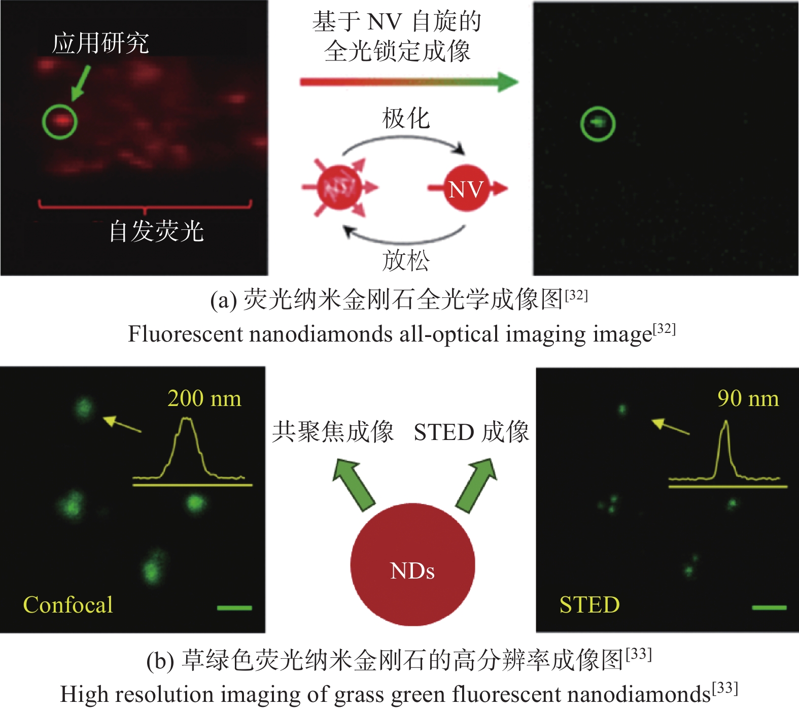
 点击查看大图
计量
- 文章访问数: 13
- HTML全文浏览量: 4
- PDF下载量: 1
- 被引次数: 0
 邮件订阅
邮件订阅 RSS
RSS